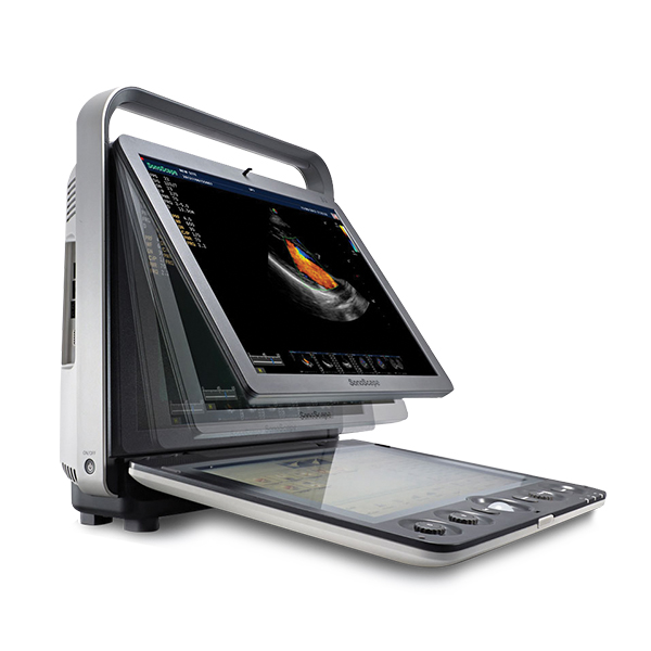

SonoScape S2

SonoScape S2

Comprehensive Functions for Streamlined Veterinary Exams

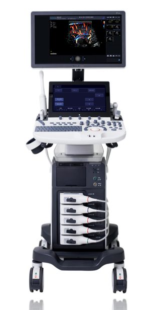
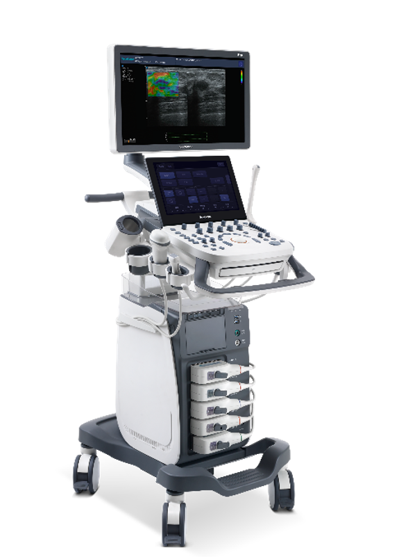
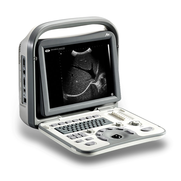
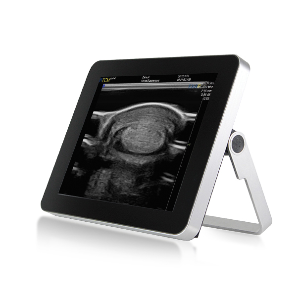
The S2 is a color Doppler system with a practical design and stable performance that goes beyond your expectations, but not your budget.

With advanced features usually found only in higher-end systems the S2 is the perfect system for general practices that require enhanced image quality, but only have basic Doppler needs. The image optimization features of the S2 keep exam times down and image quality high.
The unit is focused on bringing you the best images possible. Designed with an expansive digital beam former to provide exceptional image quality, resulting in crystal-clear images which enable you to make a confident diagnosis on a wide variety of patients.
Complete working modes, outstanding 2D performance, sensitive blood flow imaging, and 4D imaging technology give the S2 a class-leading feature set.

Upgraded Workflow, Smart Data Management

Intuitive user interface: clear menu-driven interface designed for convenient workflow.

Define your own work-style: fully customisable work settings for data table/graph, function keys, icons, and measurement formulas.

Clipboard function: quick image review during scanning.

Smart data management: retrieve, review and report with easy access to image and data storage.

Features/Specs

Advanced Features

15″ High Resolution Monitor

QWERTY Keyboard

Rechargeable Lithium Battery

Two Transducer Sockets

Ergonomic Cart (Optional)

Optimized Workflow

DICOM 3.0 Connectivity

Compound Imaging

Trapezoidal Imaging

4D Imaging (transducer dependent)

Automatic Tissue Optimization Technology (ATO)and M-Tuning (1-button scanning) deliver ease-of-use and time savings by automatically adjusting parameters to display the optimal image quality for the type of tissue being imaged.

Basic Doppler Capabilitiesincluding Color Flow,Pulsed Wave Spectral Doppler, and Power Doppler.

Compound Imagingcombines multiple coplanar images captured from different beam angles to form an integrated image.

Steerable M Modeallows for precise axis positioning, resulting in extremely accurate measurements.

Panoramic Imagingproduces an ultra-wide field of view, providing maximum information on tissue and lesions.

U-scan Technology, the latest advance in Speckle Reduction Technology, reduces noise while increasing border definition and diagnostic confidence.


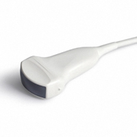
Curved Linear Probe - C344
Curved Linear – (C344) 2.0 – 6.8 MHz – Abdomen, Obstetrics, Gynaecology, Urology
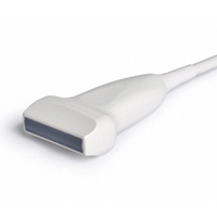
Linear Probe - L741
Linear – (L741) 4.0 – 16.0 MHz – MSK, Small Parts, Nerve, Vascular
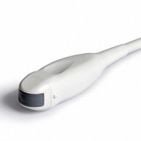
Micro-Convex Probe - C611
Micro-Convex – (C611) 3.9 – 13.0 MHz – Abdomen, Cardiac, Urology, Paediatrics

Additional information

Weight 13.6 kg